If you are worried that you have symptoms or signs of prostate cancer it is vital that you visit your GP.

What tests are there for prostate cancer?


Prostate cancer may be suspected following a digital rectal examination or blood test called PSA (Prostate Specific Antigen). However, it can only be confirmed by examining prostate tissue (a biopsy) under a microscope. Sometimes, advanced prostate cancer is diagnosed when men visit the doctor feeling unwell, with tiredness, loss of appetite and perhaps bone pain.
 

Prostate Specific Antigen (PSA) Test


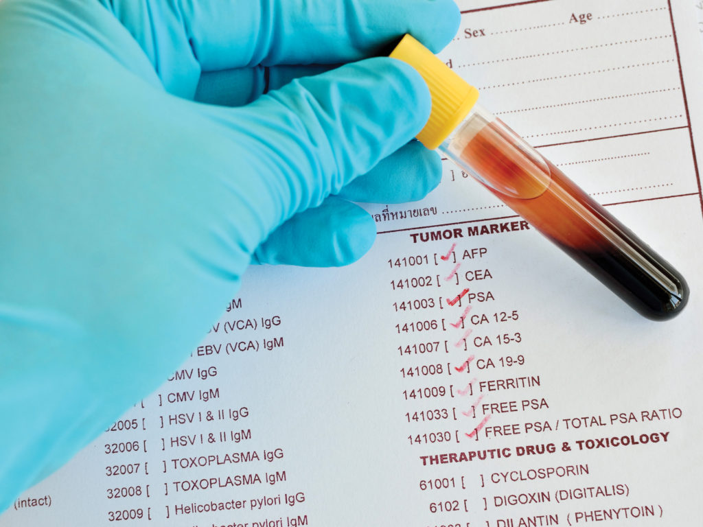
PSA is made by the prostate and some of it leaks into the blood. A small sample of blood is taken from a vein in the arm and sent to a laboratory to measure the level of PSA. You may be advised to avoid strenuous exercise and sexual activity for 48 hours before taking a PSA test as they could affect the results. It is also important to tell your doctor about any other medication or procedures you have had as these could also affect your PSA.

Only one in four men with a PSA level between 4 and 10 micrograms per litre has prostate cancer. There are various reasons for a raised PSA level. A high PSA does not necessarily mean you have cancer. Similarly,
a lower PSA does not necessarily mean you do not have cancer.

As it is generally felt that the PSA test is an insufficiently accurate indicator of prostate cancer, you may find it helpful to undergo regular PSA tests to detect any changes early. Any man over the age of 50 is entitled to a free PSA test under the NHS informed choice programme, called Prostate Cancer Risk Management.

Digital Rectal Examination (DRE)


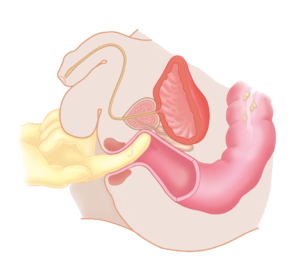
You will be asked to lie on a couch on your side with your knees drawn up towards your chin. The doctor or specialist nurse will then put a gloved finger up into your bottom. He or she will be able to feel your prostate through the rectum wall. Possible signs of prostate cancer include a prostate that feels hard or lumpy.

Further tests


Your GP will ask you to describe how you feel. Depending on the results of the digital rectal examination, urine and blood tests, your GP may refer you to a hospital for further investigation by a surgeon (urologist).

MRI (magnetic resonance imaging)


Magnetic resonance imaging scans the prostate to look for abnormal areas. If after a prostate biopsy prostate cancer is diagnosed, the scan will help determine how much cancer is present.

In most centres, a multi-parametric MRI (mpMRI) is carried out prior to prostate biopsies. If no suspicion of prostate cancer is found during the mpMRI, a patient may not need to undergo a biopsy. Other centres may be unable to carry out an mpMRI first, so will schedule MRI scans after prostate biopsies. The MRI machine is a large cylinder surrounded by a magnet.

For the scan, you will lie on a moveable table that will slide into the MRI machine for the picture of your prostate to be taken.

Likert score and PI-RAD scale


Images from the MRI are used by the radiologist to give you a Likert score. Your Likert score indicates how likely it is that you have prostate cancer and will range from 1 to 5. A score of 1 means you are very unlikely to have prostate cancer whereas a score of 5 means that you are highly likely to have prostate cancer. You could also be given a PI-RADS score, which uses a similar 1-to-5 scoring system.

Prostate biopsy


You will usually be asked to lie on your back with your knees bent and thighs apart or your legs in stirrups. An ultrasound probe is put into your back passage. A fine needle is inserted either through the area between your testicles and back passage (transperineal biopsy) or through your back passage (transrectal biopsy). The number of samples (biopsies) taken depends on your PSA level and your MRI scan report. Biopsies are usually carried out under a local anaesthetic. This means the doctor will numb the prostate, the area surrounding the prostate and the area between the back passage and the testicles (perineum). Sometimes a general anaesthetic is used. The samples are sent to the laboratory to be viewed under a microscope. You may also have a course of antibiotics in order to prevent infection.


Bone scan


A bone scan shows any spread of cancer into the bones. A tiny amount of radioactive substance is injected into your veins and taken up by abnormal bone. The images will be taken several hours after the injection. Any spread of cancer to the bones shows up as dark areas. You may also have a CT scan or MRI scan. These help to tell the difference between early and late prostate cancer.

Staging and grading

Staging and grading

Understanding treatment choices

Understanding treatment choices

Prostate Cancer Help

Prostate Cancer Help
Press enter or esc to cancel